Peripheral nerve injuries often cause sensory and motor dysfunction, which seriously affects the quality of life of patients. Although existing nerve conduits can provide physical bridging, they lack the topology, mechanical properties, and electrical signaling ability suitable for nerve growth, which makes it difficult to achieve ideal repair results. Therefore, the development of nerve conduits with bionic structure, suitable mechanical properties and electrical activity is of great significance to repair the nerve regeneration microenvironment.
Recently, Prof. Chen Huang's team from the College of Textiles, together with Dr. Yuanming Ouyang's team at the Sixth People's Hospital of Shanghai Jiaotong University School of Medicine, constructed a multifunctional nerve conduit by simultaneous loading of single-layer graphene (SLG) and nanodiamond (ND) particles in grooved polycaprolactone fibers (PCL). The catheter utilizes the grooved structure to guide the directional growth of nerve cells, and combines the mechanical enhancement of ND and the high electrical conductivity of SLG to synergistically achieve the repair of the nerve regeneration microenvironment. The above research results are summarized as ‘Anisotropic Single-layer Graphene/nanodiamond Loaded PCL Promotes Peripheral Nerve Regeneration through Mechanical-Electrical Signal Synergistic Regulation”. Conduits Provide Biophysical Cues to Manipulate Nerve Biomechanics and Bioelectric Function in the Restoration of Nerve Microenvironment’. The paper was published in Advanced Functional Materials, with Dr. Lei Zhan, a PhD student from the School of Textiles, Dr. Xu Wang, a PhD student from the Sixth People's Hospital affiliated with Shanghai Jiaotong University School of Medicine, and Master's student Yaowei Lv as the first authors.
Inspired by the anisotropic arrangement of natural nerve bundles and the force-electric properties of nerve cells, SLG/NG/PCL micron fibers were prepared by electrostatic spinning technology. The polyvinylpyrrolidone template was removed by ethanol post-treatment, and the grooved fibers were prepared by combining the solvent-induced phase separation technique with up to 14 oriented nanogrooves on the fiber surface. The specific process and principle are as follows:
High/low volatile solvents were used to dissolve PCL, and the formation of grooves during spinning could be divided into three stages:
1. high volatile solvents exchanged with water vapors;
2. water droplets evaporated to form pores, and semi-cured PCL fibers were stretched;
3. fibers were completely cured to form grooves.
The use of PVP as a sacrificial template further enhances the number and depth of grooves on the PCL fiber surface. Compared to smooth fibers, the groove structure promotes cell adhesion and migration through the oriented structure and larger specific surface area.
A two-layer catheter was prepared using a homemade device: an inner layer of doubly oriented fibers (grooves + fibers arranged in parallel) to guide nerve growth, and an outer layer of random fibers to improve mechanical properties.The addition of ND enhanced the compression modulus of the catheter by a factor of 1.6, which was attributed to the strong ND/PCL interaction and the high modulus of ND. In addition, the high electrical conductivity of the SLG itself resulted in a catheter conductivity of 0.012 S cm-¹ (Fig. 1)
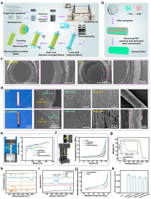
Figure 1. Preparation process and performance characterization of nerve conduits
The morphological improvement of regenerated nerves in different groups was observed by HE staining, toluidine blue (TB) staining, Sirius red (SR) staining and transmission electron microscopy (TEM). Overall, muscle atrophy was less severe in the SLG/ND/PCL group, and axon number, area, diameter, and myelin thickness were close to those of the autograft group and significantly higher than those of the control pure PCL group (Figure 2).
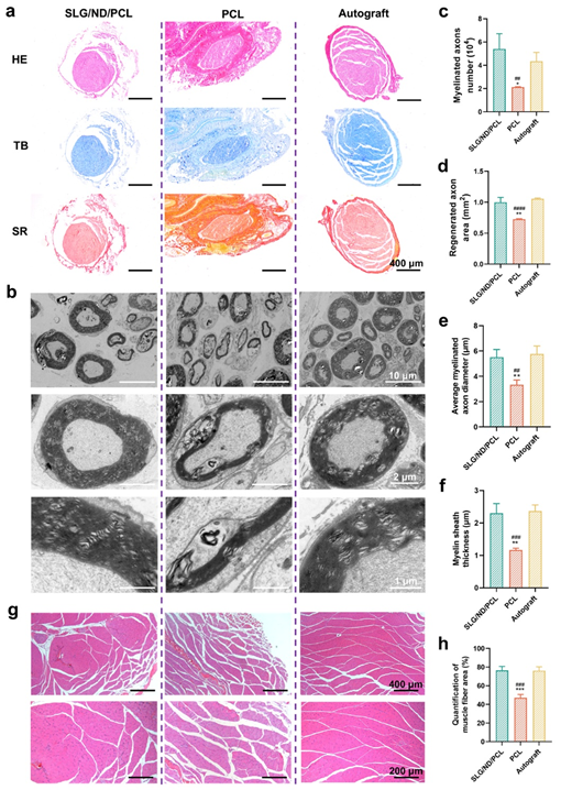
Figure 2. Expression of proteins related to nerve regeneration promoted by nerve conduit
Myelin basic protein expression was significantly enhanced in the SLG/ND/PCL group of Sherwang cells. In addition, all protein expressions related to myelin formation were downregulated after knockdown of Piezo1 gene expression, again demonstrating that the SLG/ND/PCL conduit promotes Piezo1 expression and activates related pathways to promote NFAT signaling and subsequent myelin formation in a Piezo1-dependent manner (Figure 3).
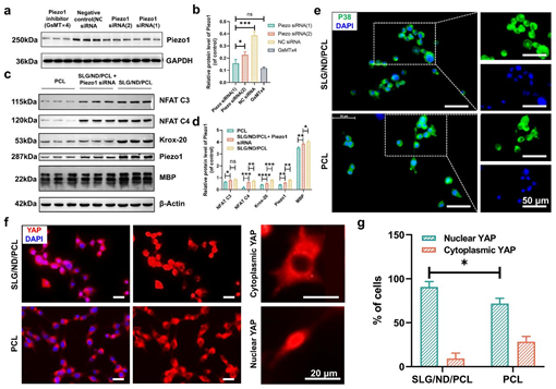
Figure 3. Mechanical deformation promotes regeneration through Piezo1 but not p38 signaling pathway
In summary, in this study, PCL fibers with controllable surface grooves were prepared and used as the building blocks of nerve conduits.The addition of SLG and ND further improved the mechanical and electrical conductivity of the nerve conduits, and synergistically promoted peripheral nerve regeneration through Piezo1-dependent synergistic effects on the growth of chervonia cells and myelin sheath formation in a significant way.
This research work was supported by the National Natural Science Foundation of China, the Natural Science Foundation of Shanghai Municipality, the Special Funds for Basic Research Operating Costs of the Central Universities, and the National Key Laboratory of Advanced Fiber Materials.
Link to full article: https://advanced.onlinelibrary.wiley.com/doi/10.1002/adfm.202419411